Idiopathic Intramedullary Osteosclerosis
Authors:
Syed A. A. Rizvi, PhD, MS, MBA
Hampton University School of Pharmacy, Hampton, VirginiaZafar Qureshi, MD; Javeria Waseem, MBBS; Rodel Reyes, MD; and Mohammad N. Manzoor, BS
UHI CommunityCare Clinic, Miami, FloridaMileydis Alonso, MS-IV
Nova Southeastern University, Fort Lauderdale, FloridaCitation:
Rizvi SAA, Qureshi Z, Waseem J, Reyes R, Manzoor MN, Alonso M. Idiopathic intramedullary osteosclerosis. Consultant. 2019;59(8):250-251.A 15-year-old boy presented with persistent pain in the mid left leg for the past 3 weeks. The pain was associated with a limp and sometimes awoke him from sleep. The patient’s mother noted swelling in his left leg with increased warmth. The patient was taking ibuprofen or acetaminophen at night to help him sleep, and she denied any history of surgery or trauma in the boy.
Physical examination. The patient was afebrile with stable vital signs. The patient’s blood pressure was 115/60 mm Hg, heart rate was 83 beats/min, respiratory rate was 22 breaths/min, height was 159 cm, and weight was 60.3 kg, for a body mass index of 23.85 kg/m2. Physical examination findings were significant for mild edema and exquisite point tenderness to palpation over the midshaft area of the left lower leg. Range of motion was normal. All other examination findings were noncontributory.
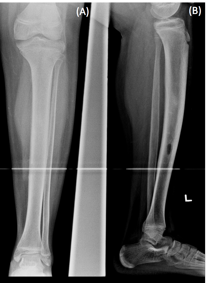
Diagnostic tests. Laboratory test results were unremarkable. Radiography findings were abnormal (Figure); thus, the case was discussed with a pediatric orthopedist, and magnetic resonance imaging (MRI) with and without intravenous contrast was obtained.

Figure. Anterior (A) and lateral (B) view radiographs of the left tibia showing intramedullary osteosclerosis and endosteal thickening.The MRI findings revealed bone marrow signal alteration within the left mid to distal tibial diaphysis. The alteration was concerning for osteomyelitis, with an irregular intramedullary area of thick peripheral-rim nodular enhancement and central hypoenhancement measuring 3.2 × 1.1 × 1.1 cm. These findings were concerning for intramedullary abscess.
The patient underwent an incisional biopsy of the left tibia, and the sample was sent for pathology examination. The lesion consisted of preexisting cancellous bone and marrow with abundant reactive woven bone formation, edema, and fibrosis. Some of the woven bone had prominent osteoblastic rimming. There was no neoplasm, infection was ruled out by negative cultures, and the changes were consistent with idiopathic intramedullary osteosclerosis.