
Cardiogenic Shock Secondary to Viral Myocarditis
An almost 3-year-old girl with a benign medical history was referred by her pediatrician to the emergency department (ED) because of concerns about possible pneumonia. The pediatrician reported that the patient had been normotensive despite tachycardia and tachypnea; she was also “clammy,” panting, and intermittently grunting. She was afebrile.
The patient had an episode of mild nausea, emesis, and diarrhea 6 days earlier that had resolved. Her mother reported that since then, the child had been lethargic, sleepy, and generally “not herself.” The child’s appetite had markedly decreased and her skin was cold and pale.
On arrival in the ED, the child was normotensive. She was in moderate respiratory distress and was grunting and moaning. She was pale, diaphoretic, and had perioral cyanosis despite normal oxygen saturation (pulse oximetry reading on room air, 100%). She had poor perfusion with cold extremities, and a delayed capillary refill (greater than 5 seconds). Her respiration rate was 50 to 60 breaths per minute.
Within 30 minutes of her arrival at the ED, the child’s health had deteriorated to the point that her blood pressure could no longer be measured without Doppler ultrasonography. Tachycardia (160 beats per minute) persisted despite fluid resuscitation with nearly 50 mL/kg of normal saline and pressor support with an epinephrine drip. External jugular veins were prominent.
Cardiac examination revealed no gallop, no muffled heart sounds, and no murmur. There was no ankle edema.
The child’s liver was initially palpable 4 cm below the costal margin. After fluid resuscitation, empiric antibiotic therapy, and airway control, her liver was palpable 7 to 8 cm below the costal margin.
Laboratory test results were as follows: white blood cell count, 18,300/μL; hemoglobin, 12.3 g/dL;platelet count, 317,000/μL; erythrocyte sedimentation rate (ESR), 6 mm/h (normal, 0 to 20 mm/h); C-reactive protein level, 0.1 mg/L (normal, 0.0 to 0.6 mg/L); electrolyte panel, normal; blood urea nitrogen, 26 mg/dL; creatinine, 0.6 mg/dL (normal, 0.2 to 0.6 mg/dL); aspartate aminotransferase, 88 U/L (normal, 5 to 60 U/L); alanine aminotransferase, 63 U/L (normal, 7 to 35 U/L); creatine kinase–MB fraction, 3.2 ng/mL (normal, 0.0 to 4.9 ng/mL); and cardiac troponin I level, 0.48 ng/mL (normal, 0.00 to 0.29 ng/mL).
Chest film as shown.
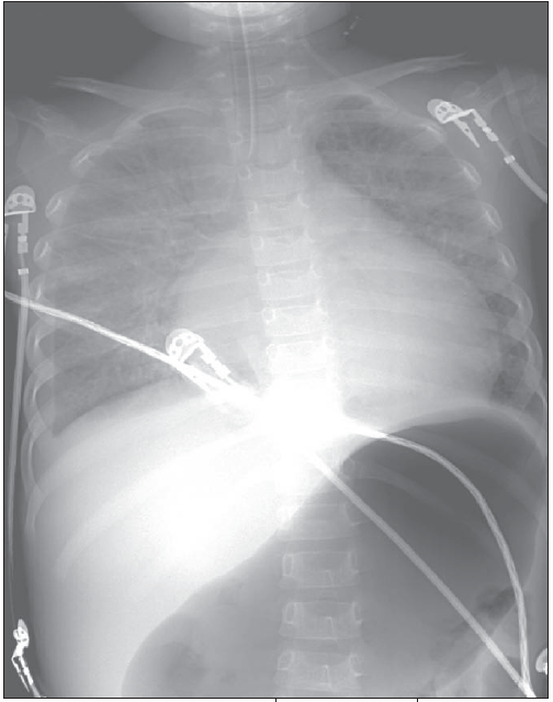
Figure – X-ray film shows endotracheal tube at tip of carina, cardiomegalay, patchy densities, and increased air in stomach.
All physical findings in this child were the result of cardiogenic shock secondary to viral myocarditis. Myocarditis usually results from viral infections. The most common are those caused by Enteroviruses (ie, coxsackievirus), adenoviruses, and influenza virus.
Manifestations of myocarditis are nonspecific. A child may initially present with flu-like symptoms or gastroenteritis. Within 1 to 4 weeks, fever, malaise, shortness of breath, tachypnea, pallor, and poorly perfused cool extremities may develop. Infants may appear critically ill, with poor feeding, fever, grunting respirations, and listlessness; symptoms of congestive heart failure are usually present. Sinus tachycardia (especially that unresponsive to fluids), jugular venous distention, weak pulses, cyanosis, poor perfusion, and hepatosplenomegaly are often found.
Chest films show cardiomegaly, interstitial pulmonary edema, or an engorged pulmonary venous pattern. In this patient’s case, the chest film showed an endotracheal tube at the tip of the carina, cardiomegaly, patchy densities, and increased air in the stomach.
An echocardiogram typically shows global cardiac enlargement with poor ventricular contractility. The white blood cell count, ESR, and C-reactive protein levels may be elevated but are nonspecific.
 Acute management includes diuretics and inotropic support for heart failure. The use of corticosteroids and immunosuppressive agents is still controversial. Mortality approaches 35% and transplantation may be needed for patients with end-stage cardiomyopathy.
This unfortunate child required extracorporeal membrane oxygenation support. She subsequently had a cerebrovascular accident. Three months later, she was still dependent on an assisted ventricular device in the pediatric ICU and was awaiting transplantation.