
Can You Identify This Periorbital Papule?
AUTHORS:
Stephanie Juliet Campbell, DO; Schield Wikas, DO; and Monte Fox, DO
CITATION:
Campbell SJ, Wikas S, Fox M. Can you identify this periorbital papule? Consultant. 2017;57(2):112.
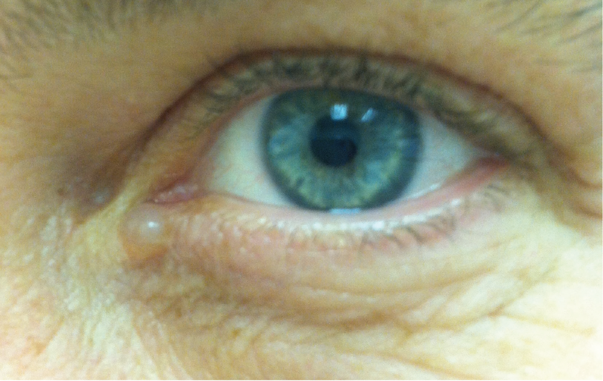
A 45-year-old woman presented with an asymptomatic solitary papule located in her left periorbital region. The lesion had been present for approximately 5 years and had remained stable in size since its onset.
Her medical history and family history were noncontributory. Physical examination revealed a single, 3-mm, flesh-colored, dome-shaped, translucent, cystic papule, with a slightly blue tint, located adjacent to the medial canthus of the left lower eyelid. The papule was sessile and was soft but tense on palpation.

What is this woman’s asymptomatic eyelid lesion?
- Chondroid syringoma
- Milial cyst
- Apocrine hidrocystoma
- Basal cell carcinoma
- Cutaneous myxoid cyst
Answer on next page
Answer: Apocrine Hidrocystoma
Apocrine hidrocystomas, also known as Moll gland cysts, cystadenomas, or sudoriferous cysts, are relatively rare benign cystic adenomas of the sweat gland and arise from the apocrine secretory coil.1 The lesions were first described in 1964 by Mehregan.2 Hidrocystomas are divided into apocrine and eccrine categories based mainly on histopathologic morphology. Even though their features are very similar, slight clinical differences exist. Apocrine hidrocystomas tend to be larger (3-15 mm) and solitary, whereas eccrine hidrocystomas may be smaller (1-6 mm) and more numerous.1,3
Epidemiology. Apocrine hidrocystomas are more common in the middle aged to elderly population, although hidrocystomas have been reported in the pediatric population as a very rare occurrence. There is a slight female predominance. There is no predilection for race.4
Pathogenesis. Apocrine hidrocystomas are thought to arise from an adenoma of the sweat gland coils, whereas eccrine hidrocystomas are thought to occur as a result of blockage of the sweat duct, causing fluid retention and dilation of the cyst. Accordingly, eccrine hidrocystomas may enlarge with heat or exercise or regress with cooler temperatures, whereas apocrine hidrocystomas usually do not change with temperature.1,3
Although the diagnosis is made clinically, histopathologic examination findings can confirm the clinical suspicion. A hidrocystoma can be a single or multilocular cyst and is lined by 2 layers of epithelium.4 The inner layer is cuboidal or columnar, and the outer layer is lined by myoepithelial cells.5,6 A notable feature of apocrine hidrocystomas is “decapitation secretion,” in which the apical cytoplasm of the glandular cell is pinched off and released into the lumen.4,5 Eccrine hidrocystomas typically lack myoepithelial cells and papillae, which distinguishes them from their apocrine counterparts.4,5
Clinical manifestations. Apocrine hidrocystomas have been described as translucent or flesh-colored, bluish dome-shaped papules that are asymptomatic but may be exacerbated by heat or exercise and regress with colder temperatures.1,7,8 The lesion is typically small but can range from 3 to 15 mm in size and may slowly grow over time.
Hidrocystomas may occur as a solitary lesion (Smith type) or multiple lesions (Robinson type).1,3 Most commonly, the lesion occurs on the head and neck, particularly on the upper or lower eyelid region or the malar area. Rare cases of hidrocystoma on the scalp, shoulders, chest, extremities, penis, vulva, ear, or lip, or within a nevus sebaceous, have been reported.3,6,9
Multiple hidrocystoma papules are associated with certain syndromes, including Schöpf-Schulz-Passarge syndrome and focal dermal hypoplasia (also called Goltz-Gorlin syndrome).8 Schöpf-Schulz-Passarge syndrome is an autosomal recessive disorder whose characteristic features are hidrocystoma of the bilateral upper and lower eyelids in conjunction with palmoplantar hyperkeratosis, hypodontia, and hypotrichosis.8 Multiple periocular hidrocystomas are one of the many cardinal features of focal dermal hypoplasia, along with microcephaly, microphthalmia, cognitive impairment, skeletal abnormalities, and papillomas of the lip, tongue, axilla, and anus.1,7 Multiple hidrocystomas have also been reported in association with Graves disease.1,3
Diagnosis. Hidrocystomas are typically diagnosed clinically. Dermoscopy can be a useful tool to increase clinical diagnostic confidence. A nonpolarized dermoscopic examination of a lesion may demonstrate a homogenous bluish-purplish center surrounded by a pale halo.10 Biopsy may be required for a definitive diagnosis in more clinically ambiguous cases.
Treatment. A variety of therapies for hidrocystomas have been reported. Traditionally, surgical methods such as incision and drainage or dissection of the cyst and electrodesiccation of the base have been used to remove the hidrocystoma,11,12 with scarring as a potential complication. Recurrence of the cyst has been reported with incision and drainage.11 The potential for scarring may be minimized with more novel approaches such as intralesional botulinum toxin A,13 carbon-dioxide or pulsed-dye laser therapy,14 chemical ablation with trichloroacetic acid,14 and hypertonic glucose sclerotherapy.11 The use of topical atropine15 and topical scopolamine16 also has been reported, although the associated potential adverse anticholinergic effects such as pupillary dilation could lead to patients discontinuing therapy.3,8
Apocrine hidrocystomas are a benign condition with a low potential for malignant transformation. As such, primary care providers can relieve the patient’s anxiety and prevent unnecessary consultations or procedures, unless the lesion is symptomatic or impedes the patient’s visual field. Our patient was not cosmetically concerned about the lesion and chose not to have it treated.
Stephanie Juliet Campbell, DO; Schield Wikas, DO; and Monte Fox, DO, are dermatologists at Tri-County Dermatology in Cuyahoga Falls, Ohio.
The lead author discusses the case in a podcast here.
References:
- Stone MS. Cysts. In: Bolognia JL, Jorizzo JL, Schaffer JV, eds. Dermatology. Vol 2. 3rd ed. Philadelphia, PA: Elsevier Saunders; 2012:1817-1828.
- Mehregan AH. Apocrine cystadenoma: a clinicopathologic study with special reference to the pigmented variety. Arch Dermatol. 1964;90(3):274-279.
- Sarabi K, Khachemoune A. Hidrocystomas—a brief review. MedGenMed. 2006;8(3):57.
- Elston DM. Sweat gland neoplasms. In: Elston DM, Ferringer T, eds. Dermatopathology. 2nd ed. Philadelphia, PA: Elsevier Saunders; 2014:86-104.
- Cysts, sinuses, and pits. In: Patterson JW. Weedon’s Skin Pathology. 4th ed. Philadelphia, PA: Churchill Livingstone Elsevier; 2016:510-529.
- Tejaswi C, Rangaraj M, Karthikeyan K. Apocrine hidrocystoma arising from nevus sebaceous on the scalp. Indian Dermatol Online J. 2016;7(2):111-113.
- Srivastava D, Taylor RS. Appendage tumors and hamartomas of the skin. In: Goldsmith LA, Katz SI, Gilchrest BA, Paller AS, Leffell DJ, Wolff K, eds. Fitzpatrick’s Dermatology in General Medicine. Vol 1. 8th ed. New York, NY: McGraw-Hill; 2012:1337-1362.
- Ovhal AG, Deshkulakarani SV, Abhange RS, Birare SD. Rare benign cystic lesions on face: apocrine hidrocystoma. Indian J Dermatol. 2016;61(2):237.
- Anzai S, Goto M, Fujiwara S, Da T. Apocrine hidrocystoma: a case report and analysis of 167 Japanese cases. Int J Dermatol. 2005;44(8):702-703.
- Duman N, Duman D, Sahin S. Pale halo surrounding a homogeneous bluish-purplish central area: dermoscopic clue for eccrine hidrocystoma. Dermatol Pract Concept. 2015;5(4):43-45.
- Osaki TH, Osaki MH, Osaki T, Viana GA. A minimally invasive approach for apocrine hidrocystomas of the eyelid. Dermatol Surg. 2016;42(1):134-136.
- Squires JA, Fish FS III. Excision, draining, and exteriorization techniques. In: Robinson JK, Hanke CW, Siegel DM, Fratila A, eds. Surgery of the Skin: Procedural Dermatology. 2nd ed. Philadelphia, PA: Mosby Elsevier; 2010:177-188.
- Bordelon JR, Tang N, Elston D, Niedt G, Lazic Strugar T. Multiple apocrine hidrocystomas successfully treated with botulinum toxin A [published online October 26, 2016]. Br J Dermatol. doi:10.1111/bjd.14753
- Anandasabapathy N, Soldano AC. Multiple apocrine hidrocystomas. Dermatol Online J. 2008;14(5):12.
- Armstrong DK, Walsh MY, Corbett JR. Multiple facial eccrine hidrocystomas: effective topical therapy with atropine. Br J Dermatol. 1998;139(3):558-559.
- Clever HW, Sahl WJ. Multiple eccrine hidrocystomas: a nonsurgical treatment. Arch Dermatol. 1991;127(3):422-423.