
Skin Disorders in Older Adults: Cutaneous Ulcers, Part 2
ABSTRACT: The most common ulcer on the lower back and buttocks is the decubitus ulcer (also known as the pressure sore, or bedsore). Most of these ulcers occur in elderly patients, particularly those who reside in long-term care facilities. Decubitus ulcers are categorized in 4 stages based on their depth of penetration. Because the recurrence rate is high, the best treatment for these ulcers is prevention. Coma blisters also result from pressure on the skin. Treatment involves removal of pressure from the affected site; the blister should be left intact. A number of infectious, vesiculobullous, and neoplastic processes can mimic decubitus ulcers, particularly herpes simplex, herpes zoster, and candidal infection, which can appear in the gluteal area.
Key words: cutaneous ulcer, ulceration, decubitus ulcer, pressure sore, bedsore, herpes simplex, herpes zoster, candidal infection
Cutaneous ulcers result from a variety of neoplastic, vascular, inflammatory, physical, and infectious processes. In part 1 of this 2-part article (CONSULTANT, August 2011), I discussed ulcers that typically occur on the legs. In this article, I focus on those that develop on the lower back and buttocks.
The most common type of ulcer in this region is the decubitus ulcer (also known as the pressure sore, or bedsore). Other ulcerative processes include herpes and neoplasms.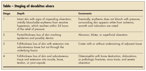


 DECUBITUS ULCERS
Decubitus ulcers develop as a result of chronic pressure and the tissue ischemia that the pressure causes. Patients who are exposed to uninterrupted pressure in excess of capillary filling pressure (approximately 32 mm Hg) over bony prominences exhibit cutaneous ulceration and necrosis.
Epidemiology. Most decubitus ulcers occur in elderly patients. The prevalence among patients in long-term care facilities is about 17% to 28%. Among those hospitalized with an acute illness, the incidence ranges from 3% to 11%. In patients with neurological impairment, the annual incidence of decubitus ulcers is about 5% to 8%.1
Staging. Decubitus ulcers are categorized in 4 stages based on their depth of penetration (Table).1 Keep in mind that a patient can present with multiple decubitus ulcers in various stages (Figure 1).
In stage 1, the skin is intact but shows signs of impending ulceration. Pressure creates initially blanchable erythema from reactive hyperemia, which resolves within 24 hours of the relief of pressure. Eventually, the erythema does not blanch with pressure, and the surrounding skin appears white from ischemia (See Figure 1). Warmth and induration are present.
Stage 2 ulcers are characterized by partial-thickness loss of skin involving the epidermis and possibly the dermis. They appear as abrasions, blisters, or superficial ulcerations (Figure 2).
Stage 3 ulcers are craters that may be associated with undermining of adjacent tissue. There is full-thickness loss of skin with extension into subcutaneous tissue but not through the underlying fascia (Figure 3).
In stage 4, full-thickness loss of skin and subcutaneous tissue occurs, and there is extension into the muscle, bone, tendon, or joint capsule. Clinical findings may include osteomyelitis with bone destruction, dislocations, or pathologic fractures; sinus tracts; and severe ulceration.
Treatment. Because the recurrence rate is higher than 90%,2 the best treatment for decubitus ulcers is to prevent them by turning patients and using special air mattresses. Stage 2 ulcers are treated with a variety of dressings, particularly hydrocolloid dressings. Stage 3 and stage 4 ulcers require surgical intervention.
COMA BLISTERS
Coma blisters are similar to decubitus ulcers in that they result from pressure applied to the skin. They differ slightly from decubitus ulcers because the pressure that generates coma blisters is more intense but of shorter duration.
Causes. Coma blisters were first described in patients who were comatose as a result of an overdosage of drugs or carbon monoxide. Drugs that have been implicated include barbiturates, methadone, hydrocodone, diazepam, amitriptyline hydrochloride, clorazepate dipotassium, meprobamate, imipramine hydrochloride, acetyl-bromo-diethylacyl-carbamide, and glutethimide.3
Clinical and histopathologic features. Coma blisters are limited to cutaneous involvement and are most commonly observed over bony eminences; however, they can occur on any part of the body. Histopathologic examination reveals intraepidermal or subepidermal vesicles with epidermal eosinophilic necrosis. The most striking and significant change is eccrine sweat gland necrosis. Similar clinical and histopathologic features can also be observed in patients with non–drug-induced coma blisters.
Like decubitus ulcers, coma blisters can sometimes appear as urticarial plaques. Such plaques, however, demonstrate eccrine duct necrosis.
Treatment. Treatment involves removal of pressure from the affected site; the blister should be left intact. When the blister breaks, an erosion or ulceration is revealed (Figure 4). Coma blisters and ulcerations sometimes occur on the ear (Figure 5). Healing occurs without scarring over a period of 2 to 4 weeks.4
OTHER CAUSES OF
ULCERATIONS ON THE
BACK AND BUTTOCKS
A number of infectious, vesiculobullous, and neoplastic processes can mimic decubitus ulcers, particularly herpes simplex (Figure 6), herpes zoster (Figure 7), and candidal infection, which can appear in the gluteal area. If you suspect that the cause of an ulcer is infectious, order a Tzanck smear, direct immunofluorescence assay, and potassium hydroxide preparation. Pemphigus can also cause erosions and ulcerations that can be confused with other types of ulcers; biopsy is needed to make the diagnosis.
Neoplastic processes can result in ulcers on any part of the body. Basal cell carcinoma commonly manifests as an ulceration, which is also known as a rodent ulcer. Any non-healing ulcer must be biopsied whether it is on the back or any other part of the body. ■
1. Bluestein D, Javaheri A. Pressure ulcers: prevention, evaluation, and management. Am Fam Physician. 2008;78:1186-1194.
2. Bates-Jensen BM, Guihan M, Garber SL, et al. Characteristics of recurrent pressure ulcers in veterans with spinal cord injury. J Spinal Cord Med. 2009;32:34-42.
3. Dunn C, Held JL, Spitz J, et al. Coma blisters: report and review. Cutis. 1990;45:423-426.
4. Waring WS, Sandilands EA. Coma blisters. Clin Toxicol (Phila). 2007;45:808-809.