
Peer Reviewed
Popsicle Panniculitis
Authors:
Maureen DeWitt, MD
Resident Physician, Children’s Hospital of Michigan, Detroit
Deepak Kamat, MD, PhD
Professor of Pediatrics and Vice Chair for Education, Wayne State University School of Medicine; Designated Institutional Official, Children’s Hospital of Michigan, Detroit
Citation:
DeWitt M, Kamat D. Popsicle panniculitis. Consultant. 2019;59(5):160.
A 3-month-old girl presented to our office for a facial rash. Her parents had noticed the rash on her face the morning of presentation.
They denied pacifier use or laying the infant in a position that would cause the patches to develop. The parents denied the use of any new soaps or lotions or exposure to pets. There had been no recent change in the infant’s diet. The lesions were not pruritic. There were no associated symptoms such as fever, vomiting, or diarrhea. She had normal appetite and activity. Findings of a review of systems were otherwise negative.
Physical examination revealed an erythematous lesion measuring 2 cm in diameter, on the left cheek and a similar lesion measuring 1 cm in diameter on the right cheek (Figure). The lesions were indurated and warm but not tender. The remainder of physical examination findings were unremarkable.
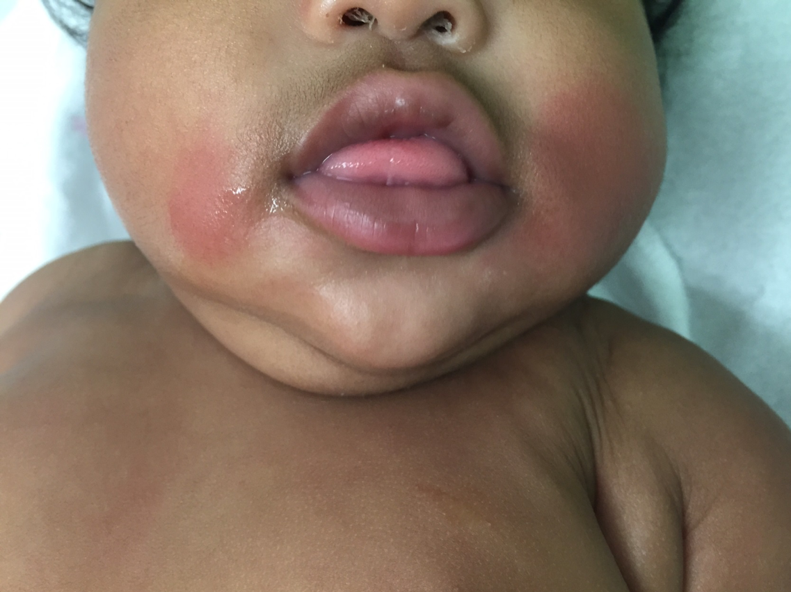
When the parents were asked whether the child had recently come in contact with anything cold, the mother remembered that she had allowed the child to chew on a wrapped popsicle the previous day. The diagnosis of popsicle panniculitis, otherwise known as cold panniculitis, was made.
Discussion. Cold panniculitis presents as erythematous plaques or subcutaneous nodules on areas that are exposed to cold. The lesions can be painful.1 The term popsicle panniculitis refers specifically to lesions on the chin or cheeks of an infant or child after sucking on a popsicle or ice cube.2 Cold panniculitis can occur hours after cold exposure or even up to 3 days after. Other examples of cold panniculitis include sites of contact with cooling blankets or icepacks, and on the thighs or buttocks of equestrians (also called equestrian panniculitis).2,3 Cold panniculitis can also occur after exposure of unprotected skin, especially the face, to wind and cold weather.4 Cold panniculitis is most commonly seen in young infants but also can occur in children and even adults.
Cold panniculitis occurs when subcutaneous fat crystalizes due to cold exposure. Unsaturated fats crystalize at colder temperatures than saturated fats. Infants have more saturated fats than older children and adults do. Because of their increased saturated fats, infants will experience cold panniculitis formation after a shorter duration of cold exposure than will adults and older children.2,3
The diagnosis is usually made clinically.4 Biopsy is generally not needed to make the diagnosis. However, if the lesion were to be biopsied, results would show a mostly lobular panniculitis with an infiltrate of lymphocytes and histiocytes, as well as a superficial and deep perivascular dermal infiltrate without vasculitis.2
There is no specific treatment for cold panniculitis. Patients should be advised to avoid the offending agent.2 The lesions generally resolve after a few weeks to a few months.1 Any hyperpigmentation associated with the lesion usually takes longer to resolve.1
This patient was seen the next month for a well-child examination, and the cold panniculitis had resolved without any residual hyperpigmentation.
References:
- Paller AS, Mancini AJ. Vasculitic disorders. In: Paller AS, Mancini AJ. Hurwitz Clinical Pediatric Dermatology: A Textbook of Skin Disorders of Childhood and Adolescence. 4th ed. Philadelphia, PA: Elsevier Saunders; 2011:chap 2
- Polcari IC, Stein SL. Panniculitis in childhood. Dermatol Ther. 2010;23(4):356-367.
- Patel AR, Husain S, Lauren CT, Garzon MC. Circular erythematous patch in a febrile infant. Pediatr Dermatol. 2012;29(5):659-660.
- Quesada-Cortés A, Campos-Muñoz L, Díaz-Díaz RM, Casado-Jiménez M. Cold panniculitis. Dermatol Clin. 2008;26(4):485-489.
1. Duncan WC, Freeman RG, Heaton CL. Cold panniculitis. Arch Dermatol. 1966;94:722-724.
2. Lemez L. Beitrag zur pathogenese der subcutanen fettgewebsnekrose neugeborener (sog. Sclerodermia neonatorum) an der hand einer kaltereaktion des subcutanen fettgewebes bei neugeborenen und jungen sauglingen. Z Kinderheilk. 1928;46:323-369.
3. Hirsch J. Fatty acid patterns in human adipose tissue. In: Handbook of Physiology. Baltimore: Williams and Wilkins; 1965:181-190.
4. Collins HA, Stahlman M, Scott HW Jr. The occurrence of subcutaneous fat necrosis in an infant following induced hypothermia used as an adjuvant in
cardiac surgery. Ann Surg. 1953;138:880-885.
5. Requena L, Sánchez Yus E. Panniculitis. Part II. Mostly lobular panniculitis. J Am Acad Dermatol. 2001;45:325-361.