
Peer Reviewed
A Nodular Lesion on the Helix of a Boy’s Ear
Authors:
Alia Abbas, BS
MD Candidate, School of Medicine, University of Mississippi Medical Center, Jackson, Mississippi
Barbara Wilson, MD
Associate Professor, Department of Dermatology, University of Virginia, Charlottesville, Virginia
Citation:
Abbas A, Wilson B. A nodular lesion on the helix of a boy’s ear. Consultant. 2019;59(5):135-136.
A 4-year-old boy presented with an asymptomatic, firm, yellow-tan nodule on the left posterior helix (Figure). The lesion had appeared shortly after birth and had continued to enlarge until approximately 2 months of age, after which its size had remained stable. He had no other medical problems and had normal development. He had no family history of similar lesions.
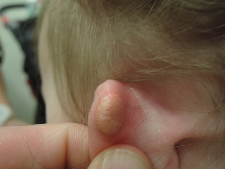
Figure. A firm yellow-tan nodule on the left posterior helix.
Answer: Juvenile Xanthogranuloma
The boy was given a clinical diagnosis of juvenile xanthogranuloma (JXG).
JXG is the most common type of non–Langerhans cell histiocytosis. It most often develops in the first year of life and more rarely appears in early adulthood.1 JXG has a predilection for boys. It is characterized by yellow to red-brown, firm, rubbery nodules measuring approximately 0.5 to 2 cm, usually arising on the head and neck.1 Most lesions are solitary, but cases of multiple nodules have also been reported.2
JXG is thought to be caused by an abnormal reactive proliferation of histiocytes in response to an unknown stimulus.1 It is not associated with lipid or metabolic abnormalities as are other xanthomatous disorders. While the condition is most likely to be benign and asymptomatic, JXG has the potential to involve internal organs and cause serious complications.3 While rare, the most common site of extracutaneous involvement is the eye. The eyes should be evaluated in any patient with JXG, and when unilateral exophthalmos is present in an infant, intraocular JXG should be considered.1
If the lesions are present with café au lait spots or a known history of neurofibromatosis type 1, there is an increased risk of an aggressive form of leukemia called juvenile monomyelocytic leukemia. Blood cell counts should be closely monitored in these patients.1,4 With the exception of cases with systemic involvement, excision is a definitive treatment5; however, lesions tend to regress on their own by age 6.1
References:
- Hernandez-Martin AD, Baselga E, Drolet BA, Esterly NG. Juvenile xanthogranuloma. J Am Acad Dermatol. 1997;36(3 pt 1):355-367.
- Dehner LP. Juvenile xanthogranulomas in the first two decades of life: a clincopathologic study of 174 cases with cutaneous and extracutaneous manifestations. Am J Surg Pathol. 2003;27(5):579-593.
- Szczerkowska-Dobosz A, Kozicka D, Purzycka-Bohdan D, Biernat W, Stawczyk M, Nowicki R. Juvenile xanthogranuloma: a rare benign histiocytic disorder. Postepy Dermatol Alergol. 2014;31(3):197-200.
- Paulus S, Koronowska S, Fölster-Holst R. Association between juvenile myelomonocytic leukemia, juvenile xanthogranulomas and neurofibromatosis type 1: case report and review of the literature. Pediatr Dermatol. 2017;34(2):114-118.
- Janssen D, Harms D. Juvenile xanthogranuloma in childhood and adolescence: a clinicopathologic study of 129 patients from the Kiel Pediatric Tumor Registry. Am J Surg Pathol. 2005;29(1):21-28.