
Peer Reviewed
Isolated Macrodactyly
AUTHORS:
Taylor A. Merritt1 • Carolyn G. Carter, MD2
AFFILIATIONS:
1University of Florida College of Medicine, Gainesville, Florida
2Department of Pediatrics, University of Florida College of Medicine, Gainesville, Florida
CITATION: Merritt TA, Carter CG. Isolated macrodactyly. Consultant. 2020;60(6):e7. doi:10.25270/con.2020.03.00023
Received November 13, 2019. Accepted March 5, 2020.
DISCLOSURES: The authors report no relevant financial relationships.
CORRESPONDENCE: Carolyn G. Carter, MD, 7046 Archer Rd, Gainesville, FL 32608 (cartcg@shands.ufl.edu)
A 5-year-old girl with a history of premature birth presented to her primary care pediatrician for her annual well-child check. At presentation, the child was normotensive and afebrile, with a body mass index (BMI) in the 98th percentile.
On physical examination, an elongated, swollen, crooked left middle finger was noted (Figure 1). The child denied any pain or loss of function at the site. Review of systems and physical examination findings were otherwise normal, aside from a failed hearing test. A referral to a pediatric orthopedics specialist was placed for management of the finger deformity.
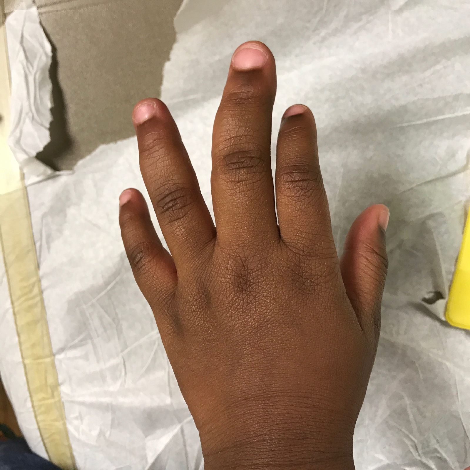
Figure 1. The patient’s enlarged left middle finger at initial presentation.
At the orthopedic visit, the child’s mother stated that she had first noticed the finger deformity when the child was 2 years of age. Since then, the child’s mother believed that the left middle finger had gotten progressively larger. The patient again denied any pain, numbness, or loss of function. On physical examination, the left middle finger and ring finger were noted to have circumferential and longitudinal enlargement, with angular deformity of the left middle finger. Complete range of motion, normal pulses, and normal sensation were noted on physical examination. No lesions, lacerations, or instability were noted.
A radiograph of the left hand demonstrated diffuse enlargement of the middle and ring fingers in a skeletally immature patient (Figure 2). There was no evidence of acute fracture, dislocation, or other osteoarticular abnormalities on the radiograph.
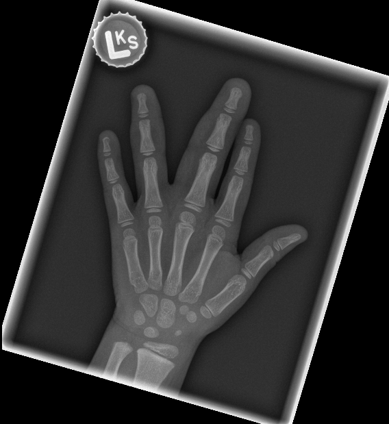
Figure 2. Radiograph of the left hand showing diffuse enlargement of the middle and ring fingers.
Based on the initial physical examination and radiographic findings, the patient was diagnosed with isolated macrodactyly of the left middle and ring fingers. Both static and progressive macrodactyly were considered as initial potential diagnoses; however, progressive macrodactyly was suspected based on the lack of digital enlargement at birth and the mother’s report of the deformity worsening over time. No immediate action was taken, and the patient was instructed to follow up regularly with the pediatric orthopedics practice.
During a physical examination with the orthopedics practice 1 year after the initial diagnosis, circumferential and longitudinal enlargement of the left middle and ring fingers with angular deformity of the left middle finger was again noted (Figure 3). Progressive longitudinal enlargement was suspected based on physical examination findings from previous visits.
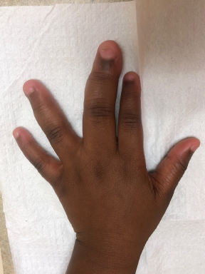
Figure 3. Enlarged left middle and ring fingers at a 1-year follow-up visit.
A second radiograph of the left hand taken at this time demonstrated a similar appearance to the first radiograph. No definitive diagnosis of progressive or static macrodactyly was made. No surgical intervention was deemed necessary, given the patient’s intact functionality and lack of significant growth of the left middle and ring fingers on the radiographic images. The patient was instructed to continue to follow up with the orthopedics practice and was informed of the possibility of surgical procedures in the future if the macrodactyly were to progress.
Approximately 2 years after her first orthopedic evaluation, the child had continued to have full range of motion and function in her fingers and hand without any further interventions (Figure 4).

Figure 4. Enlarged left middle and ring fingers at a 2-year follow-up visit.
DISCUSSION
Macrodactyly is an extremely rare disease characterized by fibro-fatty enlargement of one or multiple digits on the hands or feet in the distribution of a major peripheral nerve. This condition can present as isolated or syndromic macrodactyly. Syndromic macrodactyly, such as macrodactyly in the context of Proteus syndrome, is diagnosed when the digital enlargement is associated with other pathologies.1 Additionally, macrodactyly can be classified as static or progressive.2 Static macrodactyly, or macrodactyly simplex congenita, is characterized by digital enlargement that is present at birth and grows at the same rate as the other digits. On the other hand, progressive macrodactyly, or macrodystrophia lipomatosa, is not present at birth and grows disproportionately over time.
Generally, this condition is diagnosed based on the presenting phenotype alone. The exact etiology is still largely unknown; however, a somatic gain-in-function mutation in PIK3CA, a component of the mechanistic target of rapamycin (mTOR) signaling pathway, has been identified in several cases of isolated macrodactyly.3 This mutated gene has been linked to several overgrowth disorders, including macrodactyly, which have been classified as PIK3CA-related overgrowth spectrum (PROS). Clinical trials investigating mTOR inhibitors as a potential treatment option for PROS and progressive overgrowth disorders are ongoing but have shown a modest reduction in growth in early clinical trials.4
Macrodactyly treatment is challenging. Current treatment involves nonoperative and operative interventions. Nonoperative observation is appropriate in cases where the digital enlargement is static, limited to one or two fingers, and not hindering functionality. If the digital enlargement is progressing, operative interventions are typically necessary to control growth, reduce size, and maintain mobility. Common surgical options include debulking to decrease width, epiphysiodesis to delay longitudinal growth, nerve decompression to relieve symptoms, osteotomies to correct angulation, and amputation to assist with severe cases.5 The overall prognosis for this disease varies based on the specific subtype, but typically, one or more surgical procedures are required to treat the stiffness, immobility, and nerve compression that may result from macrodactyly.6
REFERENCES:
- Krengel S, Fustes-Morales A, Carrasco D, Vázquez M, Durán-McKinster C, Ruiz-Maldonado R. Macrodactyly: report of eight cases and review of the literature. Pediatr Dermatol. 2000;17(4):270-276. doi:10.1046/j.1525-1470.2000.01773.x
- Barsky AJ. Macrodactyly. J Bone Joint Surg Am. 1967;49(7):1255-1266.
- Rios JJ, Paria N, Burns DK, et al. Somatic gain-of-function mutations in PIK3CA in patients with macrodactyly. Hum Mol Genet. 2013;22(3):444-451. doi:10.1093/hmg/dds440
- Parker VER, Keppler-Noreuil KM, Faivre L, et al. Safety and efficacy of low-dose sirolimus in the PIK3CA-related overgrowth spectrum. Genet Med. 2019;21(5):1189-1198. doi:10.1038/s41436-018-0297-9
- Cerrato F, Eberlin KR, Waters P, Upton J, Taghinia A, Labow BI. Presentation and treatment of macrodactyly in children. J Hand Surg Am. 2013;38(11):2112-2123. doi:10.1016/j.jhsa.2013.08.095
- Ezaki M, Beckwith T, Oishi SN. Macrodactyly: decision-making and surgery timing. J Hand Surg Eur Vol. 2019;44(1):32-42. doi:10.1177/1753193418796441