
Peer Reviewed
A Case of Oral Tori in a 60-Year-Old Woman
Authors:
Tejaswi Marri, BS; Cameron Dodd, BA; and Lynnette Mazur, MD, MPH
McGovern Medical School at the University of Texas Health Science Center, Houston, Texas
Citation:
Marri T, Dodd C, Mazur L. A case of oral tori in a 60-year-old woman. Consultant. 2020;60(2):45-47, 50. doi:10.25270/con.2020.02.00002
A 60-year-old woman presented for a routine checkup. She had no medical concerns but wanted to discuss treatment options for her oral tori (Figures 1 and 2). The bony lesions had first appeared in her 30s and had remained asymptomatic until the past 2 or 3 years. During that time, they had progressively grown and multiplied to the point that she was unable to eat without pain and/or bleeding. For the past several weeks, she had limited her diet to milkshakes and smoothies. Because of her worsening symptoms, she was referred to an oral maxillofacial surgeon for further evaluation and management.
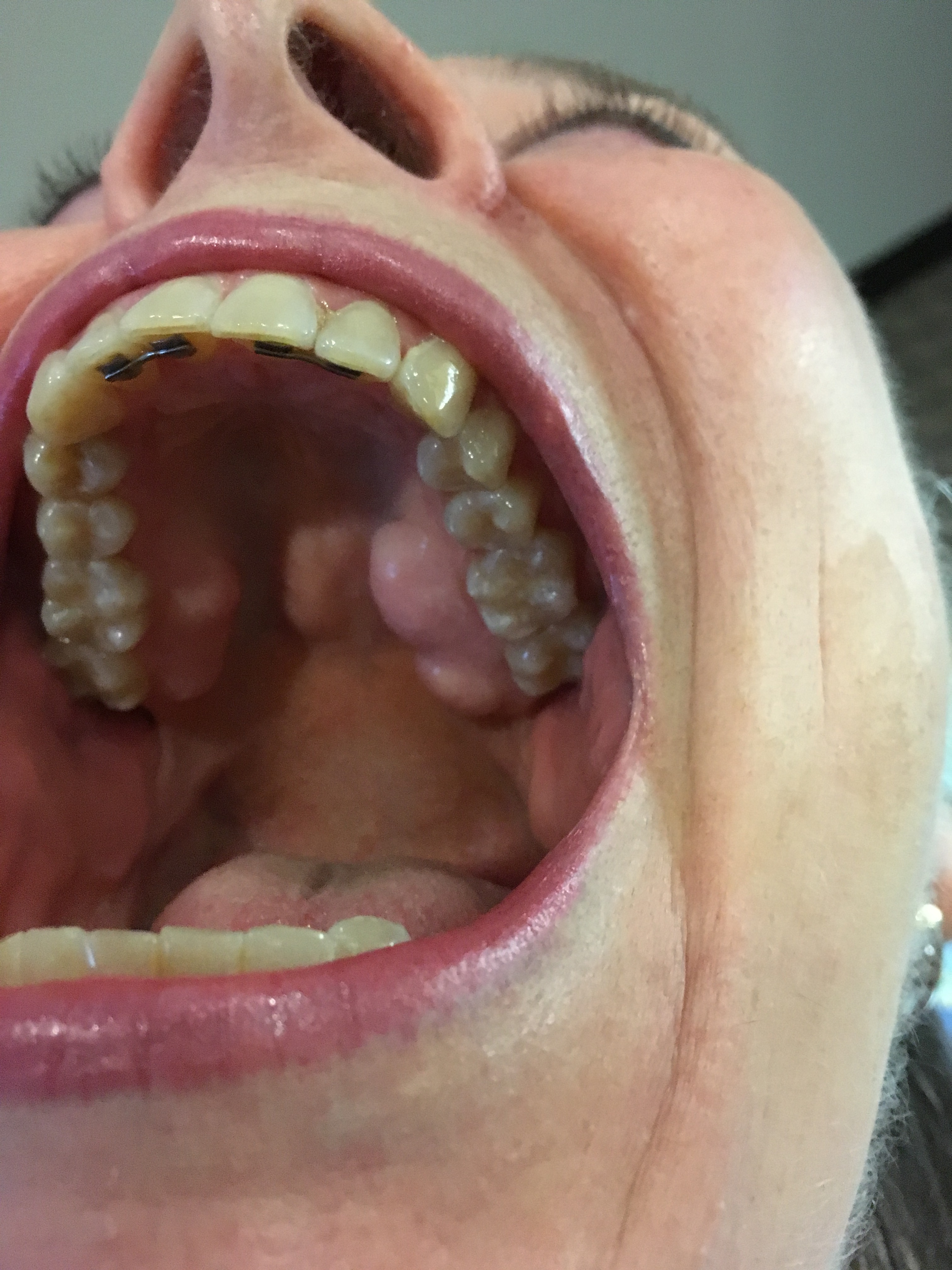
Figure 1. Torus palatinus visible along the hard palate.

Figure 2. Bilateral torus mandibularis visible along the lingual aspect of the mandible.
DISCUSSION
Oral tori are nonpathologic, nodular protuberances composed of cortical bone covered by a thin, poorly vascularized mucosa.1 The two most common forms are torus palatinus (TP) and torus mandibularis (TM).1,2 TP forms along the midline of the hard palate, whereas TMs form along the lingual aspect of the mandible and is usually bilateral.2,3 Tori typically develop during late adolescence and gradually increase in size throughout adulthood.3 When small, they rarely cause symptoms or pain and are usually an incidental finding during routine clinical or dental examinations.4
Their prevalence varies by ethnicity and geographical region, but tori are more commonly found in Eskimos, Native Americans, Norwegians, and Thais.4,5 Prevalence ranges from 12% and 14% in patients from Trinidad and Tobago, respectively, to 27% in patients from Thailand.1,6,7 In the United States, TP is the most prevalent torus, occurring in 20% of the population, while TM has a prevalence of 6%.8,9 TM is more common in men, whereas TP is more common in women.2,5,8,10 Concurrence of TP and TM ranges from 3% to 23%.1,7,11,12
Although the etiology of oral tori is unknown, genetic and environmental factors may have a role.13 Oral tori are thought to follow an autosomal dominant pattern of inheritance, and masticatory stress, masticatory hyperfunction and bruxism are thought to be risk factors.10,14 Superficial trauma in the oral cavity and lifestyle factors such as consumption of fish, a calcium-rich diet, and vitamin deficiency are also associated with their development.2,5-7,10,15 One study showed a higher bone-mineral density in patients with tori.16 There is also a correlation between tori and temporal mandibular joint dysfunction and obstructive sleep apnea.17,18
If radiologic studies are performed, radiopaque masses with a higher density than surrounding bone may be noted.4,5 Oral tori must be differentiated from other growths in the oral cavity, including ossifying fibroma, osteoma, mucocele, osteochondroma, osteoblastoma, osteosarcoma, and osteoid osteoma (Table).3,8,13 There does not appear to be a strong relationship between oral tori and other bone or hereditary exostoses. However, buccal exostoses may be associated with more serious syndromes such as Gardner syndrome and fibrous dysplasia.3,19
Table. Differential Diagnoses for Oral Tori | ||
Condition | Clinical Features | Diagnostic Features |
Ossifying fibroma20 | Benign | Radiography: Well circumscribed lesion with osteoblastic rimming |
Osteoma21 | Benign | Radiography: Well defined smooth growth protruding from other bone Histology: Dense lamellar or trabecular bone in orderly arrangement |
Osteochondroma22 | Benign | Radiography: Continuation of bone cortex and medullary cavity into bony outgrowth with calcified cartilaginous cap; cartilaginous cap best seen on computed tomography or MRI |
Mucocele23 | Benign | Diagnosed based on direct visualization of blue cystic swelling, history of trauma, and location of lesion |
Osteoblastoma21 | Benign but aggressive | Radiography: Well circumscribed lesion that can be radiolucent or have speckled mineralization; 4-6cm in size |
Osteosarcoma of the jaw24 | Malignant; type 1, unknown etiology; type 2, older patients with Paget disease, irradiation of the facial region, and fibrous dysplasia of the bone | Radiography: Periosteum elevation with sunburst appearance |
Osteoid osteoma21 | Benign | Radiography: Radiolucent osteoid core with surrounding sclerosis and mineralization |
A biopsy may be needed to distinguish oral tori from the other growths.3,4 Because they are self-limiting, benign, and typically painless, removal of tori is not warranted in most cases.5,9,13 Indications for surgical excision include esthetic concerns, disturbance of phonation, restriction of masticatory functions, sensitivity (due to thin overlying mucosa), traumatic inflammation or ulceration, retention of food particles, or to allow for proper fitting of oral prostheses.5,9,13,25 In some cases the cortical bone excised from the tori may be repurposed as a source for grafts in certain procedures.9 One study demonstrated positive results when using patients’ own tori for grafting bone defects between teeth when periodontal pockets are present.26 Tori can safely be used in place of other graft materials such as allografts, xenografts, alloplasts, and other locations of autografts.9,26
OUTCOME OF THE CASE
Because of the pain and difficulty with eating, our patient opted for surgical excision (Figure 3). Within a week after the procedure, she progressed from a liquid diet to soft foods without problem.

Figure 3. Postoperative photo of torus palatinus excision.
- Jainkittivong A, Langlais RP. Buccal and palatal exostoses: prevalence and concurrence with tori. Oral Surg Oral Med Oral Pathol Oral Radiol Endod. 2000;90(1):48-53.
- Antoniades DZ, Belazi M, Papanayiotou P. Concurrence of torus palatinus with palatal and buccal exostoses: case report and review of the literature. Oral Surg Oral Med Oral Pathol Oral Radiol Endod. 1998;85(5):552-557.
- Medsinge SV, Kohad R, Budhiraja H, Singh A, Gurha S, Sharma A. Buccal exostosis: a rare entity. J Int Oral Health. 2015;7(5):62-64.
- Ladizinski B, Lee KC. A nodular protuberance on the hard palate. JAMA. 2014;311(15):1558-1559.
- García-García AS, Martínez-González J-M, Gómez-Font R, Soto-Rivadeneira Á, Oviedo-Roldán L. Current status of the torus palatinus and torus mandibularis. Med Oral Patol Oral Cir Bucal. 2010;15(2):e353-e360.
- Al-Bayaty HF, Murti PR, Matthews R, Gupta PC. An epidemiological study of tori among 667 dental outpatients in Trinidad & Tobago, West Indies. Int Dent J. 2001;51(4):300-304.
- Bruce I, Ndanu TA, Addo ME. Epidemiological aspects of oral tori in a Ghanaian community. Int Dent J. 2004;54(2):78-82.
- Chatterjee S. Bony bumps in the mouth. Cleve Clin J Med. 2016;83(1):17-1
- Sonnier KE, Horning GM, Cohen ME. Palatal tubercles, palatal tori, and mandibular tori: prevalence and anatomical features in a U.S. population. J Periodontol. 1999;70(3):329-336.
- Loukas M, Hulsberg P, Tubbs RS, et al. The tori of the mouth and ear: a review. Clin Anat. 2013;26(8):953-960.
- Kolas S, Halperin V, Jefferis K, Huddleston S, Robinson HBG. The occurrence of torus palatinus and torus mandibularis in 2,478 dental patients. Oral Surg Oral Med Oral Pathol. 1953;6(9):1134-1141.
- Rajendra Santosh AB, Jones T, Venugopal H, et al. Prevalence of oral tori among medical and dental students at the University of the West Indies. Dentistry 3000. 2016;4(1). doi:10.5195/d3000.2016.55.
- Kannan S, Muthusamy S, Muthu K, Sidhu P. Multiple bony overgrowths in the mouth - report of two cases. Clin Cases Miner Bone Metab. 2015;12(3):260-261.
- Cortes ARG, Jin Z, Morrison MD, Arita ES, Song J, Tamimi F. Mandibular tori are associated with mechanical stress and mandibular shape. J Oral Maxillofac Surg. 2014;72(11):2115-2125.
- Eggen S. Torus mandibularis: an estimation of the degree of genetic determination. Acta Odontol Scand. 1989;47(6):409-4
- Hosoi T, Yoda T, Yamaguchi M, Amano H, Orimo H. Elderly women with oral exostoses had higher bone mineral density. J Bone Miner Metab. 2003;21(2):120-122.
- Morrison MD, Tamimi F. Oral tori are associated with local mechanical and systemic factors: a case-control study. J Oral Maxillofac Surg. 2013;71(1):14-22.
- Palm E, Franklin KA, Marklund M, et al. Mandibular tori size is related to obstructive sleep apnea and treatment success with an oral appliance. Sleep Breath. 2014;18(2):431-438.
- Chandna S, Sachdeva S, Kochar D, Kapil H. Surgical management of the bilateral maxillary buccal exostosis. J Indian Soc Periodontol. 2015;19(3):352-355.
- Khan SA, Sharma NK, Raj V, Sethi T. Ossifying fibroma of maxilla in a male child: report of a case and review of the literature. Natl J Maxillofac Surg. 2011;2(1):73-79.
- Greenspan A. Benign bone-forming lesions: osteoma, osteoid osteoma, and osteoblastoma. Clinical, imaging, pathologic, and differential considerations. Skeletal Radiol. 1993;22(7):485-500.
- Mohanty S, Gupta H, Dabas J, Kumar P. Osteochondroma of maxillofacial region: tumor arising from two different developmental bones. J Oral Maxillofac Pathol. 2016;20(2):329.
- Senthilkumar B, Mahabob MN. Mucocele: an unusual presentation of the minor salivary gland lesion. J Pharm Bioallied Sci. 2012;4(suppl 2):S180-S182.
- Chaudhary M, Chaudhary SD. Osteosarcoma of jaws. J Oral Maxillofac Pathol. 2012;16(2):233-238.
- Castro Reino O, Perez Galera J, Perez Cosio Martin J, Urbon Caballero J. Surgery of palatal and mandibular torus [in Spanish]. Rev Actual Odontoestomatol Esp. 1990;50(394):47-50, 53-56.
- Santhanakrishnan M, Rangarao S. Mandibular tori: a source of autogenous bone graft. J Indian Soc Periodontol. 2014;18(6):767-771.