
Peer Reviewed
An Atlas of Lumps and Bumps, Part 3
AUTHORS:
Alexander K. C. Leung, MD1,2 —Series Editor • Benjamin Barankin, MD3 • Joseph M. Lam, MD4 • Kin Fon Leong, MD5
AFFILIATIONS:
1Department of Pediatrics, University of Calgary, Calgary, Alberta, Canada
2Alberta Children’s Hospital, Calgary, Alberta, Canada
3Toronto Dermatology Centre, Toronto, Ontario, Canada
4Department of Pediatrics and Department of Dermatology and Skin Sciences, University of British Columbia, Vancouver, British Columbia, Canada
5Pediatric Institute, Kuala Lumpur General Hospital, Kuala Lumpur, Malaysia
CITATION:
Leung AKC, Barankin B, Lam JM, Leong KF. An atlas of lumps and bumps, part 3. Consultant. 2021;61(4):e12-e15. doi:10.25270/con.2021.03.00011
DISCLOSURES:
Dr Leung is the series editor. He was not involved with the handling of this paper, which was sent out for independent external peer review.
CORRESPONDENCE:
Alexander K. C. Leung, MD, #200, 233 16th Ave NW, Calgary, AB T2M 0H5, Canada (aleung@ucalgary.ca)
EDITOR’S NOTE:
This article is part of a series describing and differentiating dermatologic lumps and bumps. To access previously published articles in the series, visit https://bit.ly/35J1I1v.
Lymphangioma Circumscriptum
Lymphangioma circumscriptum, also known as microcystic lymphatic malformation, is a benign saccular dilatation of the cutaneous and subcutaneous lymphatics.1 The condition is usually congenital but can be acquired. The latter may result from damage to and obstruction of lymphatic vessels with subsequent lymphectasia. Acquired causes of lymphangioma circumscriptum include filariasis, tuberculosis, scrofuloderma, lymphogranuloma venereum, hidradenitis suppurativa, lichen planus, scleroderma, recurrent cellulitis, severe herpes simplex infection, Crohn disease, local surgery, and radiotherapy.2-8 Congenital lymphangioma circumscriptum usually occurs at birth or shortly thereafter, while the acquired type can occur at an age.7
Lymphangioma circumscriptum is characterized by clusters of vesicles resembling frog spawn. The color depends on the content. Whitish, yellow, or light tan coloration is due to the color of the lymph fluid, while reddish or blue coloration is due to the presence of erythrocytes in the lymph fluid as a result of hemorrhage and hemoglobin degradation (Figures 1-3).9 Typical dermoscopic features include yellow lacunae surrounded by pale septa without inclusion of blood and pink to yellow lacunae alternating with bluish or dark red lacunae due to inclusion of blood.4
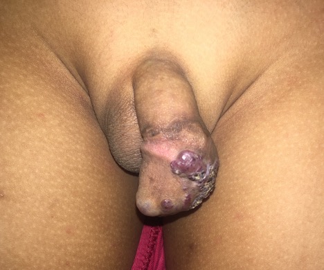
Figure 1.

Figure 2.
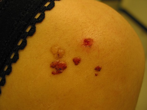
Figure 3.
Intermittent swelling, hemorrhage, or oozing of fluid from the superficial vesicles may occur.6,10 With time, the vesicles may undergo verrucous changes and have a warty appearance.1,6 The condition is usually asymptomatic but occasionally can cause pruritus or pain.6,11 Sites of predilection include the trunk, oral cavity, buttocks, axilla, proximal extremities, and less commonly, the genitalia.12,13
Pearly Penile Papules
Pearly penile papules, also known as papillae coronae glandis or papillomatosis corona penis and hirsuties coronae glandis, are benign lesions of the penis.14 Clinically, pearly penile papules present as small, smooth, soft, flesh-colored, pearly white, yellowish, pinkish, or rarely completely translucent papules (Figures 4 and 5).15,16 The papules are usually dome- or conical-shaped, although they may be filiform.17,18 The lesions are closely aggregated and range from 1 to 2 mm in diameter and 1 to 4 mm in length.17-20 They are usually uniform in size and shape and are symmetrically distributed.15,16 Typically, the papules occur in a single, double, or multiple rows circumferentially distributed on the corona and sulcus of the glans penis.15,16 The papules tend to be more prominent on the dorsum of the corona and less prominent toward the frenulum.15,16,18,21 At times, these rows may encircle the glans penis entirely.21,22 Profound proliferating papules running radially from the urethral meatus to the corona, confluent over the entire glans penis, have been described.20,23 Rarely, the papules are found on the dorsal and ventral penile shaft.20,24
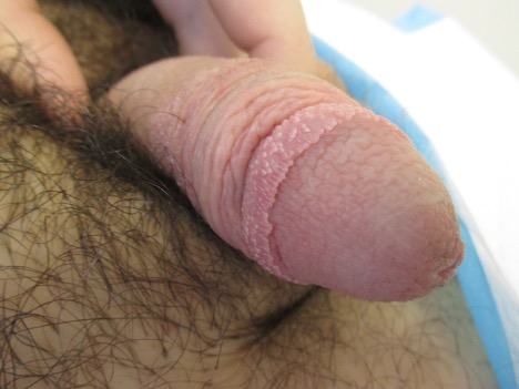
Figure 4.
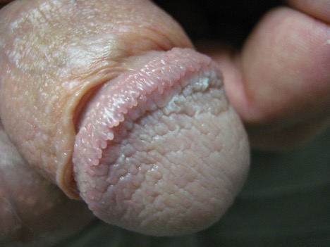
Figure 5.
Pearly penile papules usually develop in postpubertal boys, with a peak in late adolescence and early adulthood.15,16 Thereafter, the incidence decreases with age.20,25 The incidence has been estimated from 8% to 38% in adolescent boys and young men worldwide.14-16 The condition is less common among circumcised boys and more common among African American boys.14,21,25 The papules are asymptomatic and are often discovered incidentally. Pearly penile papules are benign and do not undergo malignant transformation.14 Structurally, they are related to angiofibromas.14
For someone who is familiar with the condition, it is a spot diagnosis. For someone who is not familiar with the condition, pearly penile papules are often mistaken for genital warts or sexually transmitted diseases and can lead to undue anxiety. The diagnosis can be aided by dermoscopy, which shows a whitish-pink grape-like or cobblestone pattern in a few rows with each papule containing central dotted, hairpin, or comma-like vessel structures surrounded by whitish crescent-shaped rims.14,22,26,27 Unlike genital warts, pearly penile papules do not show desquamation, which is seen as an irregular reflection on dermoscopy.14,21
References
- Adikari S, Philippidou M, Samuel M. A rare case of acquired lymphangioma circumscriptum of the penis. Int J STD AIDS. 2017;28(2):205-207. doi:10.1177/0956462416657238
- Bonini J, Ducharme O, Ponroy B, Dauendorffer JN, Baubion E. Acquired lymphangioma circumscriptum of the penis treated by electrocoagulation. Case Rep Dermatol. 2019;11(3):260-263. doi:10.1159/000503137
- Callander JA, Davies BM, Hill G. Acquired lymphangioma circumscriptum of the vulva secondary to severe herpes simplex infection. Sex Transm Infect. 2020;96(3):233-234. doi:10.1136/sextrans-2019-054224
- Dong J, Lin EY, Zhang L, Chen LQ. Dermoscopic features of cutaneous lymphangioma circumscriptum of the scrotum. Chin Med J (Engl). 2020;133(17):2126-2128. doi:10.1097/CM9.0000000000000957
- Ghalamkarpour F, Asadi-Kani Z, Moradi A, Zaresharifi S, Khazaei P. Acquired lymphangioma circumscriptum secondary to tuberculosis: a rare case report. Dermatol Ther. 2020;33(4):e13463. doi:10.1111/dth.13463
- Gude G, Gupta P, Sharma RK, Rajwanshi A. Primary lymphangioma circumscriptum of the vulva presenting as warty plaques. Australas J Dermatol. 2019;60(4):305-307. doi:10.1111/ajd.13014
- Kumar S, Mittal A, Chandna A. Penoscrotal lymphangioma circumscriptum following circumcision. Turk J Urol. 2018;44(5):437-440. doi:10.5152/tud.2018.89633
- Piernick DM 2nd, Mahmood SH, Daveluy S. Acquired lymphangioma circumscriptum of the genitals in an individual with chronic hidradenitis suppurativa. JAAD Case Rep. 2017;4(1):64-66. doi:10.1016/j.jdcr.2017.10.014
- Zaballos P, Del Pozo LJ, Argenziano G, et al. Dermoscopy of lymphangioma circumscriptum: a morphological study of 45 cases. Australas J Dermatol. 2018;59(3):e189-e193. doi:10.1111/ajd.12668
- Singh B, Hoosen K. Peri-umbilical lymphangioma circumscriptum associated with intra-abdominal lymphatic malformations. Dermatopathology (Basel). 2019;6(2):105-110. doi:10.1159/000496387
- Vatopoulou A, Anagnostopoulos A, Sujeewa F. Congenital lymphangioma circumscriptum of the vulva. J Obstet Gynaecol Res. 2019;45(10):2137-2138. doi:10.1111/jog.14086
- Ayhan E. Lymphangioma circumscriptum: good clinical response to isotretinoin therapy. Pediatr Dermatol. 2016;33(3):e208-e209. doi:10.1111/pde.12839
- Kreuter A, Oellig F, Kuntz T, et al. Acquired lymphangioma circumscriptum in high-grade penile intraepithelial neoplasia. Int J STD AIDS. Published online October 29, 2020. doi:10.1177/0956462420951094
- Love LW, Badri T, Ramsey ML. Pearly penile papule. In: StatPearls. Treasure Island (FL): StatPearls Published online October 1, 2020.
- Leung AK, Barankin B. Pearly penile papules. J Pediatr. 2014;165(2):409. doi:10.1016/j.jpeds.2014.03.019
- Leung AK, Barankin B, Leong KF, Hon KL. Penile warts: an update on their evaluation and management. Drugs Context. 2018;7:212563. doi:10.7573/dic.212563
- Gouveia AI, Borges-Costa J, Soares-Almeida L. Atypical pearly penile papules mimicking primary syphilis. Acta Dermatovenerol Croat. 2014;22(4):311-312.
- Agrawal SK, Bhattacharya SN, Singh N. Pearly penile papules: a review. Int J Dermatol. 2004;43(3):199-201. doi:10.1111/j.1365-4632.2004.02057.x
- Sapra P, Sapra S, Singh A. Pearly penile papules: effective therapy with pulsed dye laser. JAMA Dermatol. 2013;149(6):748-750. doi:10.1001/jamadermatol.2013.3130
- Yang A, Blaya Alvarez B, Makhija M, Sebaratnam DF. Profound pearly penile papules. Australas J Dermatol. 2020;61(3):280-282. doi:10.1111/ajd.13288
- Aldahan AS, Brah TK, Nouri K. Diagnosis and Management of Pearly Penile Papules. Am J Mens Health. 2018;12(3):624-627. doi:10.1177/1557988316654138
- Honigman AD, Dubin DP, Chu J, Lin MJ. Management of pearly penile papules: a review of the literature. J Cutan Med Surg. 2020;24(1):79-85. doi:10.1177/1203475419887730
- Vesper JL, Messina J, Glass LF, Fenske NA. Profound proliferating pearly penile papules. Int J Dermatol. 1995;34(6):425-426. doi:10.1111/j.1365-4362.1995.tb04445.x
- O'Neil CA, Hansen RC. Pearly penile papules on the shaft. Arch Dermatol. 1995;131(4):491-492. doi:10.1001/archderm.1995.01690160121025
- Maranda EL, Akintilo L, Hundley K, et al. Laser therapy for the treatment of pearly penile papules. Lasers Med Sci. 2017;32(1):243-248. doi:10.1007/s10103-016-2065-x
- Micali G, Lacarrubba F. Augmented diagnostic capability using videodermatoscopy on selected infectious and non-infectious penile growths. Int J Dermatol. 2011;50(12):1501-1505. doi:10.1111/j.1365-4632.2011.05087.x
- Ozeki M, Saito R, Tanaka M. Dermoscopic features of pearly penile papules. Dermatology. 2008;217(1):21-22. doi:10.1159/000118509