
Peer Reviewed
Acanthoma Fissuratum
AUTHORS:
Norhan Shamloul, BS
Drexel University College of Medicine, Philadelphia, Pennsylvania
Ronald F. Maceyko, MD
Allegheny Health Network Dermatology, Pittsburgh, Pennsylvania
CITATION:
Shamloul N, Maceyko RF. Acanthoma fissuratum [published online December 17, 2019]. Consultant360.
We report two unusual cases of acanthoma fissuratum (AF), one occurring within the concha and resulting from newer-generation hearing aids, and the other on the bridge of the nose due to the use of a continuous positive airway pressure (CPAP) device.
CASE 1
A 77-year-old man with a history of hearing loss presented to the clinic with a chief concern of a lesion on the right ear that had been present for 2 weeks. He had been referred by his primary care physician to rule out cutaneous malignancy. The patient had been wearing hearing aids for many years, with the current half-shell set having been used for the past 2 years. He stated that the lesion was irritated by his hearing aid.
On examination, a 5-mm, well-defined, erythematous, slightly edematous coffee bean–shaped papule involving the right conchal bowl and just lateral to the external auditory meatus was noted (Figure 1). Exactly one-half of this lesion was covered by his hearing aid (Figure 2).

Figure 1. A coffee bean–shaped papule on the right conchal bowl.
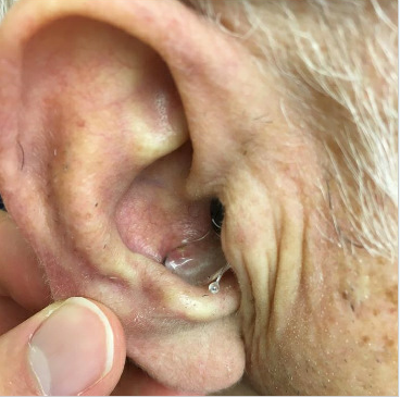
Figure 2. Hearing aid covering exactly one-half of the lesion.
Dermoscopic examination of the lesion revealed neither arborizing telangiectasia nor asymmetry of color or structure. The lesion was determined to be inflammatory in nature and was treated with intralesional triamcinolone acetonide injection, 0.5 mg.
Follow-up examination 2 weeks later revealed the same lesion (now measuring 6 mm) conforming to the area where the hearing aid abutted the skin. A clinical diagnosis of AF was established, and the patient was advised to discontinue the use of the hearing aid in the right ear to potentially avoid the need for a skin biopsy.
Follow-up examination of the right concha 4 weeks later revealed that the lesion had resolved completely (Figure 3). He was advised to have his hearing aids adjusted so they would fit better. He switched to a different set of hearing aids, after which he was free of any lesions or discomfort of the right concha.
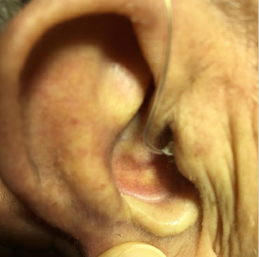
Figure 3. Complete resolution of AF 4 weeks after discontinuation of wearing the previous hearing aid
Interestingly, the patient returned to the clinic 13 months later with similar concerns, but this time of the bilateral ears. He had recently switched to new hearing aids with a plastic ear hook. Examination of each ear revealed a focal erythematous depression with surrounding mild edema of the conchal bowl where the monofilament ear hooks of the hearing aid contacted the skin (Figure 4). The stabilizer ear hook portion was displaced from the concha bowl, which provided immediate relief. Once again, he was advised to have his audiologist adjust the fit of the hearing aid, this time decreasing the length of the ear hook and ensuring the end was smooth, gently tapered, and not needlelike.
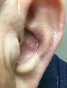
Figure 4. Early AF developing from a monofilament ear hook irritating the skin.
CASE 2
A 58-year-old man with a history of psoriasis, psoriatic arthritis, chronic obstructive pulmonary disease, and obstructive sleep apnea presented with a 1-week history of a painful lesion on the bridge of the nose. He had been wearing a CPAP mask during sleep for 3 years.
Physical examination revealed a 7-mm, firm, tender, erythematous, centrally superficially ulcerated nodule with peripheral erythema (Figure 5).
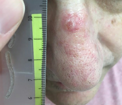
Figure 5. A lesion on the of bridge of the nose at the site of contact with a CPAP mask.
The patient preferred to have a diagnosis established with certainty as soon as possible, so a biopsy was performed to rule out malignancy. Histologic sections of the specimen demonstrated irregular epidermal acanthosis with localized scale crust present, peripheral to a central ulcer, beneath which there was a dense, suppurative infiltrate. These histologic findings, along with the clinical picture, suggested that the lesion was ulcerated AF secondary to the patient’s use of a CPAP device.
The patient was reassured about the benign nature of the lesion and was encouraged to see his respiratory therapist for adjustments to his mask. He did so, and the lesion resolved.
DISCUSSION
AF, formerly termed granuloma fissuratum, is a benign condition that may mimic cutaneous malignancy. It is characterized by focal thickening of the skin in response to minor trauma caused by trauma from chronic friction or pressure at the affected site. The term acanthoma fissuratum is preferred to granuloma fissuratum, given that histopathology examination demonstrates acanthosis and granulation tissue rather than granulomatous inflammation.
The process was first described in 1932 by Sutton, who presented 2 cases of granuloma fissuratum involving the upper labioalveolar fold secondary to ill-fitting dentures.1 In 1965, Epstein described granuloma fissuratum of the ear.2 AF is frequently observed secondary to chronic low-grade trauma from eyeglasses; this occurs on the superior ear where the temple portion of ill-fitting eyeglasses runs over the helix, and on the nasal sidewall, where the nose pads rest.2,3
Common locations for the disease process include the lateral aspect of the bridge of the nose and the retroauricular and superior auricular sulci of the ears. However, involvement of other sites has been described in the literature. AF of the posterior fourchette refers to recurrent splitting of the skin of the vulva associated with severe pain with vaginal penetration.4 AF of the penis is characterized by painful, erythematous, circumscribed plaques with a central furrow on the penile shaft caused by tight-fitting underwear.5 One other case has been reported of a person with AF secondary to a hearing aid; that lesion involved the external auditory meatus.6(p304)
AF classically presents as a unilateral firm, flesh-colored papule, plaque, or nodule with a central groove dividing the lesion into halves.1 The morphology has been described as having a coffee-bean appearance.7 Histopathology examination reveals acanthosis with hyperkeratosis and variable parakeratosis, with central epidermal attenuation of the longitudinal groove, which may contain keratin.3 The patient’s history characteristically reveals chronic trauma, usually from a device that comes into continuous direct contact with the skin. The mainstay of treatment for AF is removal of the chronic, irritating stimulus. This usually leads to resolution of the lesion within a few weeks. Other treatment modalities include intralesional corticosteroids, surgical excision, and electrosurgery in persistent cases.3
Although it is a benign condition, AF often generates a differential diagnosis to include basal cell carcinoma and squamous cell carcinoma. As such, it is important to elicit a thorough history from the patient to avoid unnecessary procedures and distress. Often, a 2- to 3-week trial of removal of the suspected offending agent (when feasible) and rechallenge allows the diagnosis to be confirmed without the need for further diagnostic or therapeutic interventions. These cases illustrate the importance of recognizing this benign entity that develops secondary to chronic friction from medical devices. Additional causes of AF are likely to be discovered as individuals wear devices chronically irritating the skin.
ADDITIONAL CONTRIBUTION:
Kathryn S. Maceyko, MSN, CRNP, FNP-BC provided manuscript review and editorial assistance.
REFERENCES:
- Sutton RJ Jr. A fissured, granulomatous lesion of the upper labio-alveolar fold. Arch Derm Syphilol. 1932;26(3):425-427. doi:10.1001/archderm.1932.01450030423004
- Epstein E. Granuloma fissuratum of the ears. Arch Dermatol. 1965;91(6):621-62 doi:10.1001/archderm.1965.01600120053012
- Deshpande NS, Sen A, Vasudevan B, Neema S. Acanthoma fissuratum: lest we forget. Indian Dermatol Online J. 2017;8(2):141-14 doi:10.4103/2229-5178.202267
- Kennedy CM, Dewdney S, Galask RP. Vulvar granuloma fissuratum: a description of fissuring of the posterior fourchette and the repair. Obstet Gynecol. 2005;105(5 pt 1):1018-1023. doi:10.1097/01.AOG.0000158863.70819.53
- Lee JI, Lee YB, Cho BK, Park HJ. Acanthoma fissuratum on the penis. Int J Dermatol. 2013;52(3):382-384. doi:10.1111/j.1365-4632.2011.04903.x
- Gonzalez SA, Moore AGN. Acanthoma fissuratum of the outer auditory canal from a hearing aid. In: Abstracts of papers presented at the 27th Annual Meeting of the American Society of Dermatopathology. San Francisco, USA. November 29–December 1, 1989. J Cutan Pathol. 1989;16(5):291-331. doi:10.1111/j.1600-0560.1989.tb00057.x
- Sand M, Sand D, Brors D, Altmeyer P, Mann B, Bechara FG. Cutaneous lesions of the external ear. Head Face Med. 2008;4:2. doi:10.1186/1746-160X-4-2