
Peer Reviewed
Diffuse Red Eruptions in a Young Boy
AUTHORS:
Oliver Wisco, DO1 • Tatiana Abrantes, BS2 • Allison Robbins, MD3AFFILIATIONS:
1Director of Cutaneous Oncology, Mohs Surgeon, Department of Dermatology, The Warren Alpert Medical School of Brown University, Rhode Island
2Medical Student, The Warren Alpert Medical School of Brown University, Providence, Rhode Island
3Resident Physician, Dermatology Department, The Warren Alpert Medical School of Brown University, Rhode IslandCITATION:
Wisco O, Abrantes T, Robbins A. Diffuse red eruptions in a young boy. Consultant. 2022;62(8):e13-e15. doi:10.25270/con.2021.11.00009Received July 5, 2021. Accepted July 9, 2021. Published online November 19, 2021.
DISCLOSURES:
The authors report no relevant financial relationships.CORRESPONDENCE:
Oliver J. Wisco, DO, FAAD, FACMS, The Warren Alpert Medical School of Brown University, 593 Eddy Street, APC, 10th Floor, Providence RI 02903 (Oliver_wisco@brown.edu)A 10-year-old boy presented to our clinic with his parents with a 6-month history of a diffuse rash on his trunk and extremities (Figures 1-3). The rash had developed abruptly, and individual lesions were characterized as pruritic and occasionally burned.
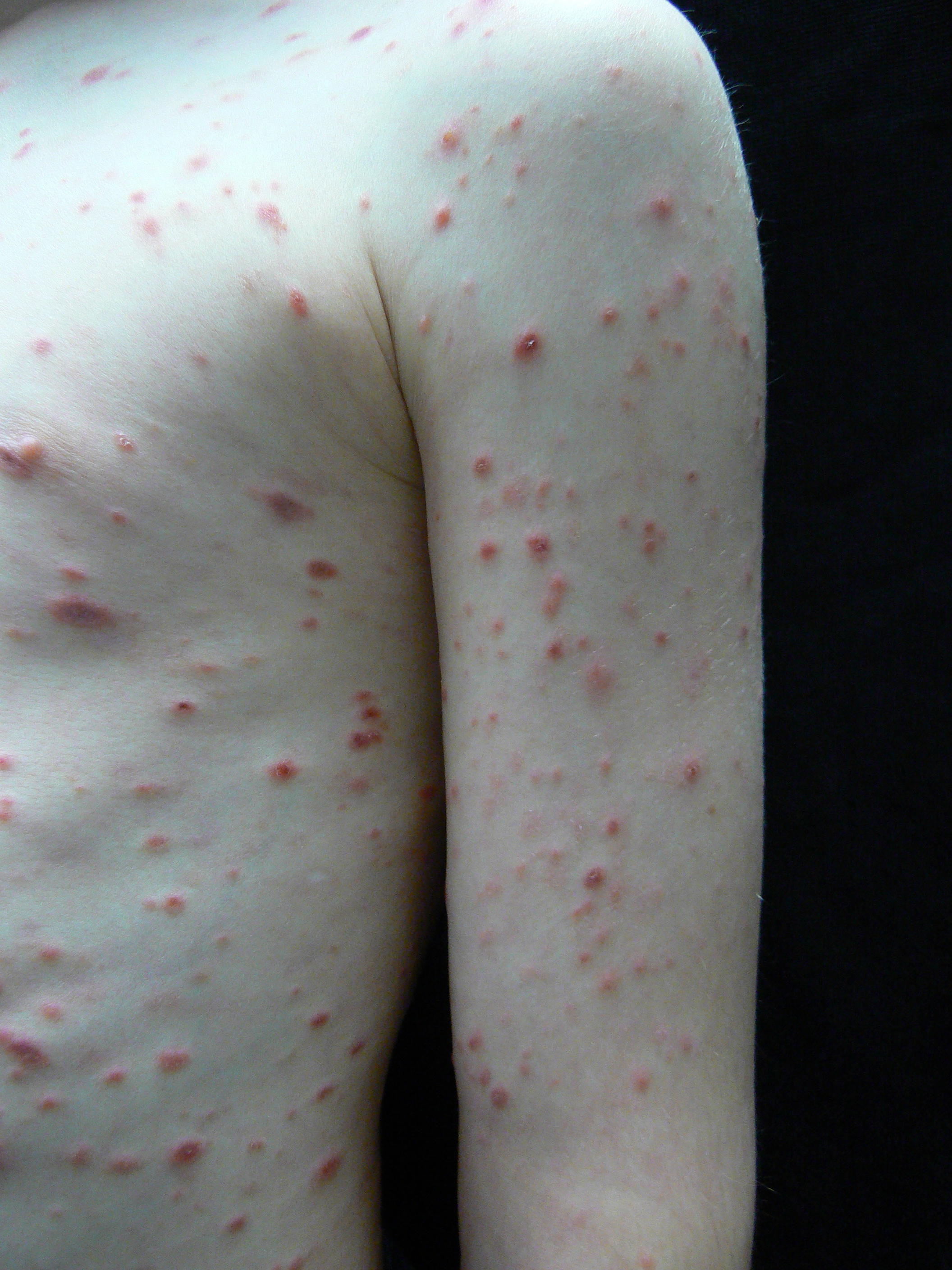
Figure 1. A diffuse rash was noted over the left side of the patient.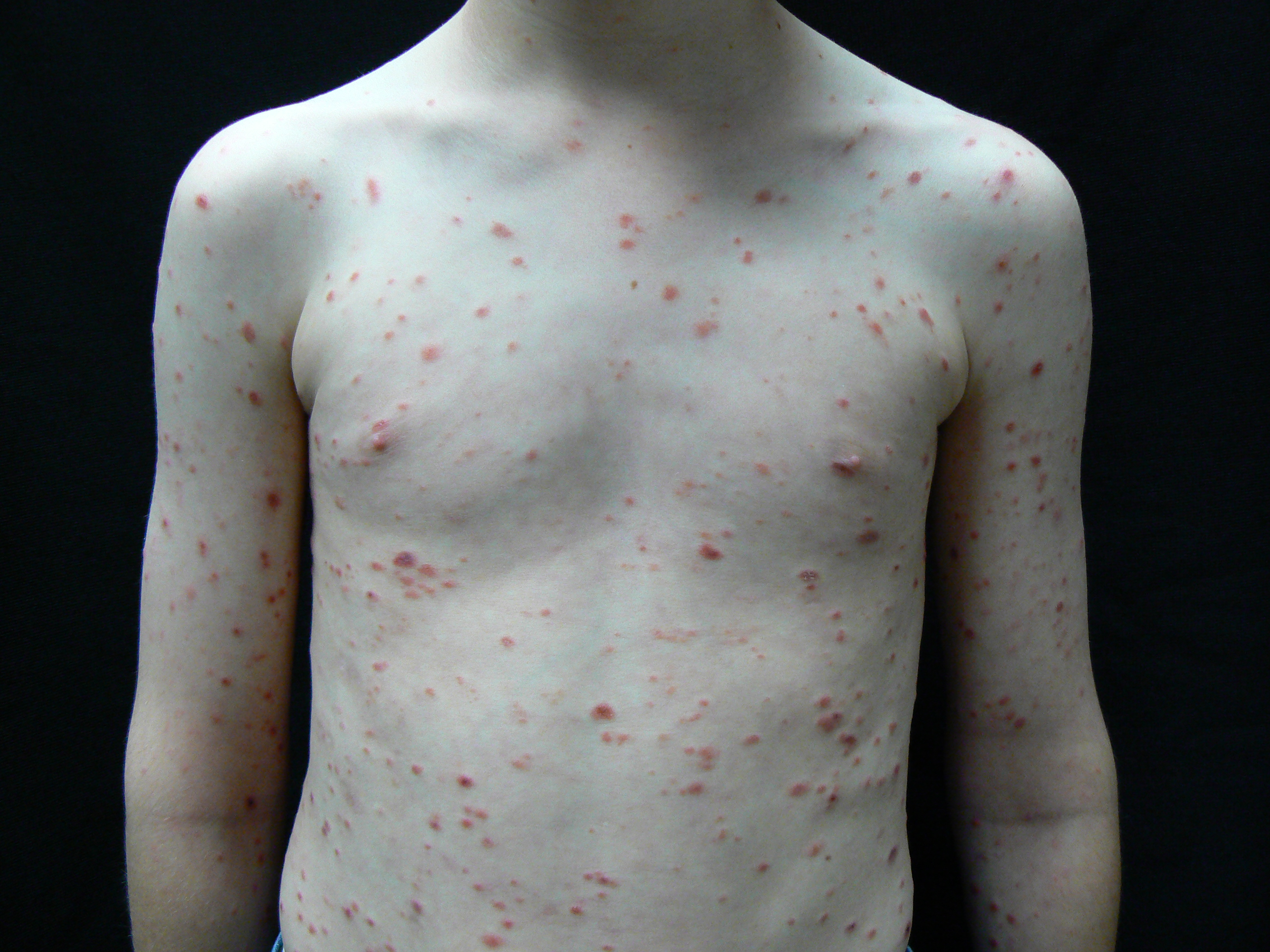
Figure 2. A diffuse rash was noted over the patient's torso, upper extremities, and shoulders.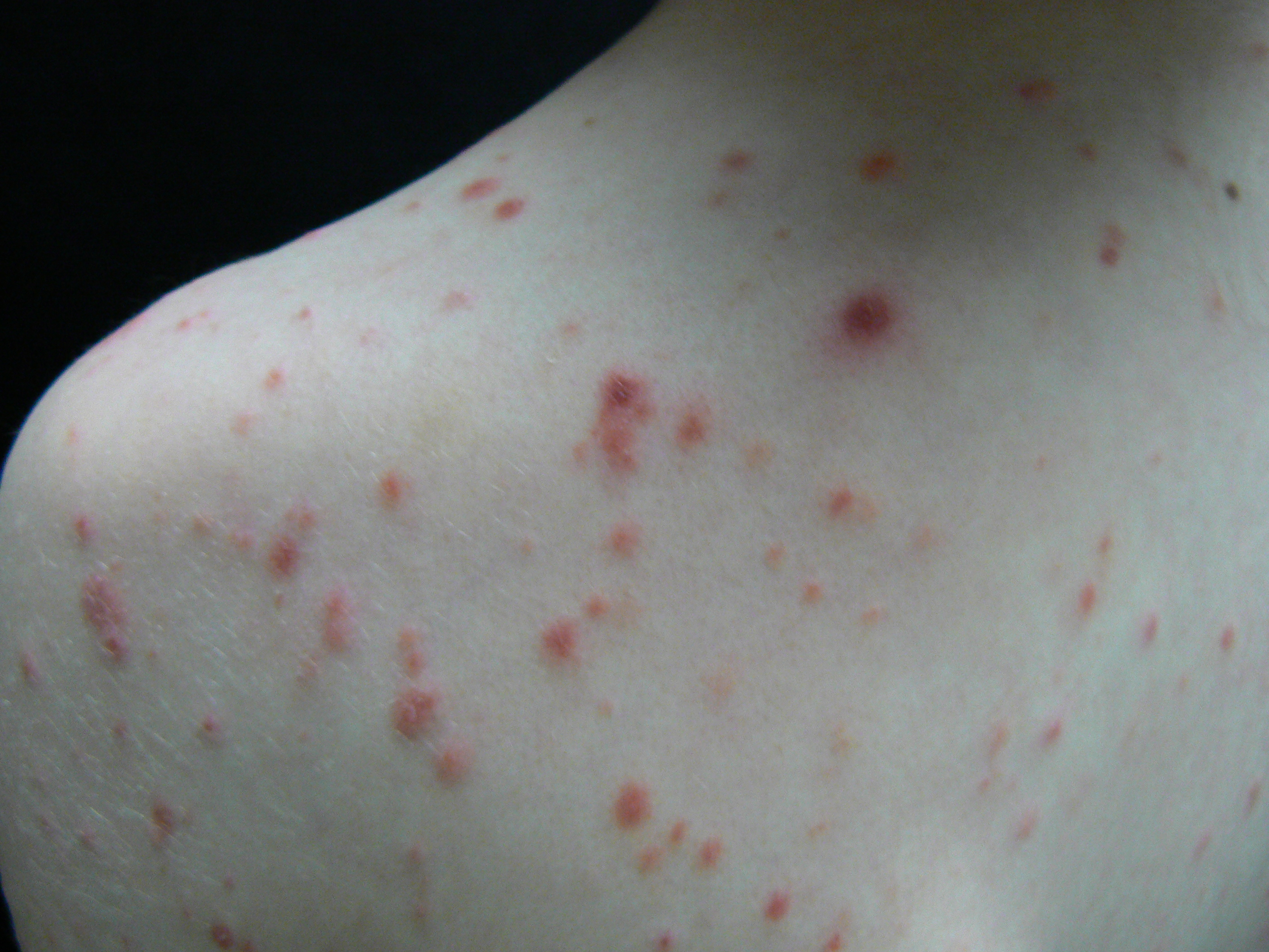
Figure 3. A diffuse rash was noted over the patient's shoulders.Prior to presentation at our clinic, previous attempts to treat the rash included erythromycin, 250 mg, 3 times daily for 4 weeks, without success, followed by clobetasol, 0.05%, ointment daily for 2 weeks. Despite treatment, new lesions continued to arise. The patient was otherwise healthy.
Upon physical examination, the patient was sitting comfortably, without persistently scratching his lesions. Diffusely scattered across his entire body were reddish-brown papules and small plaques with overlying fine micaceous scale and scattered crust.
Answer and discussion on next page.
1. Wood GS, Reizner GI. Other papulosquamous disorders. In: Bolognia J, Schaffer JV, Cerroni L, eds. Dermatology. 4th ed. Elsevier; 2017:161-174.
2. Zang JB, Coates SJ, Huang J, Vonderheid EC, Cohen BA. Pityriasis lichenoides: Long-term follow-up study. Pediatr Dermatol. 2018;35(2):213-219. https://doi.org/10.1111/pde.13396
3. Pereira N, Brinca A, Manuel Brites M, José Julião M, Tellechea O, Gonçalo M. Pityriasis lichenoides et varioliformis acuta: case report and review of the literature. Case Rep Dermatol. 2012;4(1):61-65. https://doi.org/10.1159/000337745
4. Ediale C, Felix K, Anderson K, Ahn C, McMichael AJ. An atypical presentation of PLEVA: case report and review of the literature. J Drugs Dermatol. 2019;18(7):690-691. https://jddonline.com/articles/dermatology/S1545961619P0690X&download=1
5. Bellinato F, Maurelli M, Gisondi P, Girolomoni G. A systematic review of treatments for pityriasis lichenoides. J Eur Acad Dermatol Venereol. 2019;33(11):2039-2049. https://doi.org/10.1111/jdv.15813
6. Jung F, Sibbald C, Bohdanowicz M, Ingram JR, Piguet V. Systematic review of the efficacies and adverse effects of treatments for pityriasis lichenoides. Br J Dermatol. 2020;183(6):1026-1032. https://doi.org/10.1111/bjd.18977
7. Gnann JW Jr, Whitley RJ. Clinical practice. Herpes zoster. N Engl J Med. 2002;347(5):340-346. https://doi.org/10.1056/nejmcp013211
8. Moy A, Sun J, Ma S, Seminario-Vidal L. Lymphomatoid papulosis and other lymphoma-like diseases. Dermatol Clin. 2019;37(4):471-482. https://doi.org/10.1016/j.det.2019.05.005
9. Brandon A, Mufti A, Gary Sibbald R. Diagnosis and management of cutaneous psoriasis: a review. Adv Skin Wound Care. 2019;32(2):58-69. https://doi.org/10.1097/01.asw.0000550592.08674.43
10. Schadt C. Pityriasis rosea. JAMA Dermatol. 2018;154(12):1496. https://doi.org/10.1001/jamadermatol.2018.3290