
Authors:
Amana N. Nasir, MD and Neal S. Leleiko, MD, PhD
Brown University, Providence, RI
Citation:
Nasir AN, Leleiko NS. Eosinophilic esophagitis. Consultant for Pediatricians. 2007;6(3):181.
A 9-year-old boy was brought to the emergency department after complaining that a piece of chicken was stuck in his throat. He had dinner 3 hours earlier. According to the mother, he had been complaining of mid-sternal chest pain since that time. On examination, he was sitting forward, drooling, and in considerable discomfort. He had mild tachycardia (112 beats per minute), but vital signs were otherwise normal.
The patient underwent esophagogastroduodenoscopy (A), during which a piece of chicken was removed. Further examination of the esophagus revealed linear furrowing, concentric mucosal rings, and adherent white patches (B)—findings characteristic of eosinophilic esophagitis (EE).1

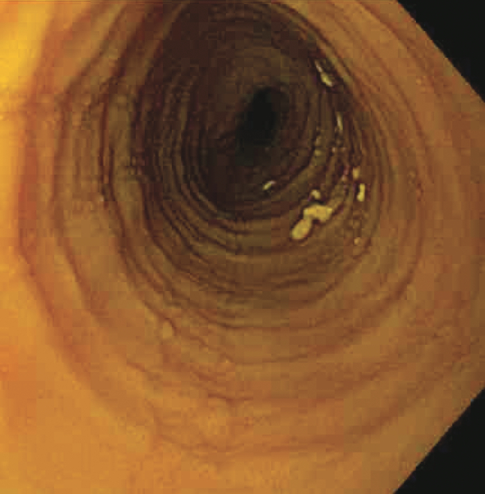
EE is increasingly being recognized as the cause of a wide spectrum of clinical manifestations, including feeding refusal, vomiting, abdominal pain, dysphagia, and food impaction. The etiology is poorly understood but has been linked to allergy: many affected patients have food and aeroallergen hypersensitivity.2 Data also support a strong familial association; nearly 10% of parents of patients with EE have a history of esophageal strictures, and about 8% of those have biopsy-proven EE.3
The number and location of eosinophils help differentiate EE from gastroesophageal reflux disease (GERD). Up to 6 eosinophils per high-power field (HPF) may indicate GERD, whereas more than 24 eosinophils per HPF appear to indicate EE. The esophageal tissue obtained from this patient during the upper GI procedure contained 40 eosinophils per HPF along with the other typical histological features of EE, including a thickened mucosa with basal layer hyperplasia and papillary lengthening.4
Treatment consists of antigen-elimination trials, anti-inflammatory agents, and antacid therapy—because gastric acid may trigger esophageal eosinophilia. If this approach is unsuccessful, a more restricted diet or an exclusive elemental (amino acid-based) formula is recommended.5
This child had experienced previous episodes of dysphagia and abdominal pain that occasionally responded to over-the-counter antacids. He had mild allergies. The child was treated with a proton pump inhibitor, once daily, and fluticasone(, twice a day via metered-dose inhaler (with instructions to swallow each puff rather than inhale). The medications were discontinued after 3 months. The patient has been symptom-free for the past 6 months.